

How a Russian surgeon turned frozen bodies into the first 3D human atlas
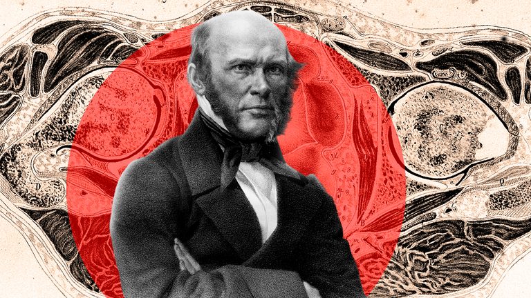
A carpenter's saw and freezing temperatures were a century ahead of computed tomography (CT) and magnetic resonance imaging (MRI).
In the mid-19th century, all anatomical textbooks were based on autopsies of fresh cadavers. But, as soon as a surgeon opened the chest or abdominal cavity, the organs shifted, losing their true outlines. Outstanding Russian surgeon Nikolai Pirogov understood that such illustrations were misleading students. This prompted him to seek a fundamentally different way to document anatomy.
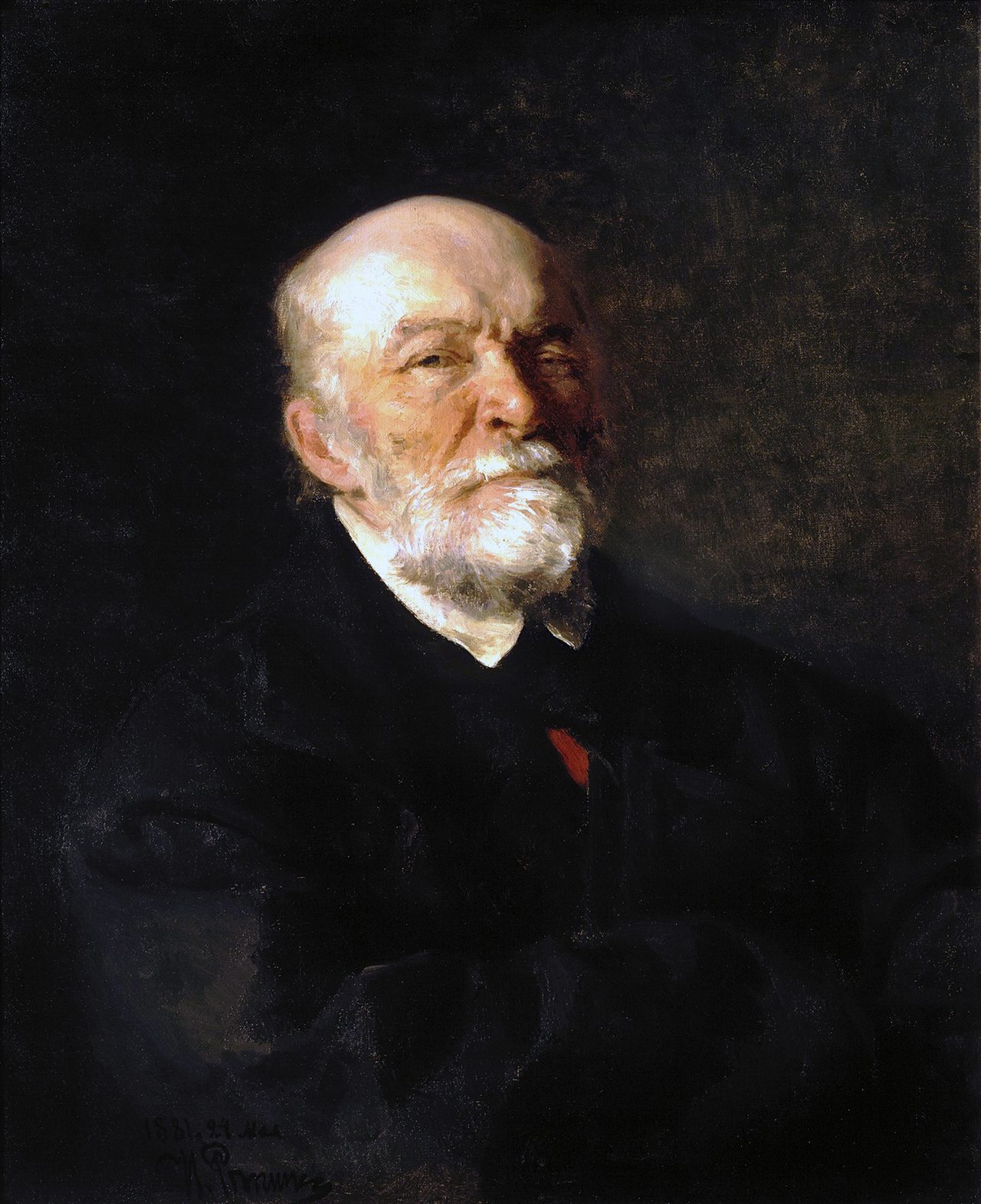
The solution was to freeze the body until it was as hard as stone and then saw it into the thinnest possible slices. Pirogov personally supervised the process: the cadavers were kept at a temperature of approximately -25°C (-13°F) for 2-3 days. The cuts were made with a mechanical saw designed to produce slices 5-10 mm thick with 6-7 mm gaps between them. To obtain a three-dimensional view, each cadaver was cut in three mutually perpendicular planes. This is how hundreds of specimens were created.
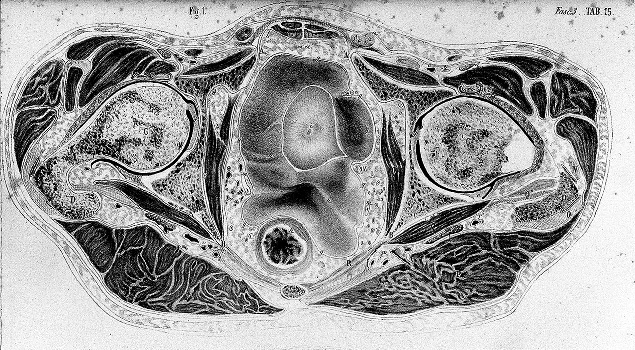
All that remained was to transfer these frozen sections onto paper. Pirogov placed glass or transparent paper, gridded into a square pattern, over the surface of each cut section. He then traced the contours of all visible structures: muscles, bones, blood vessels and internal organs. This grid served as a coordinate system, allowing the life-size image to be transferred with mathematical precision to the final sheet. By combining sections from different planes, the relative positions of organs could be reconstructed with a precision previously unattainable.
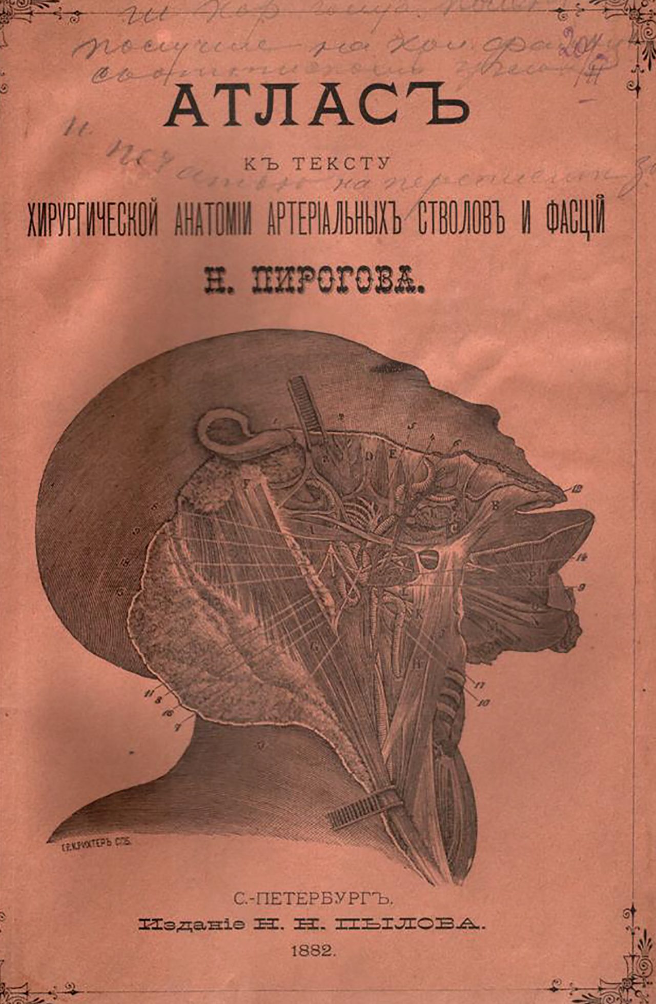
The result of many years of work was the publication ‘Topographic Anatomy, Illustrated with Sections Through the Frozen Human Body in Three Directions’, published between 1852 and 1859. It comprised four volumes, 224 tables and more than 1,000 life-size drawings. Pirogov himself proudly noted that "the position of many organs – the heart, stomach and intestines – turned out to be quite different from what is usually observed during autopsies." The atlas went on to become a standard reference for several generations of physicians worldwide.